Draw It to Know It Circle of Willis
| Circumvolve of Willis | |
|---|---|
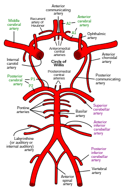 Diagram of the arterial apportionment at the base of operations of the brain (inferior view), the circumvolve of Willis is drawn in the upper half. Claret flows up to the brain through the vertebral arteries and through the internal carotid arteries. | |
| Details | |
| Identifiers | |
| Latin | Circulus arteriosus cerebri Circulus Willisii |
| MeSH | D002941 |
| TA98 | A12.two.07.080 |
| TA2 | 4516 |
| FMA | 50454 |
| Anatomical terminology [edit on Wikidata] | |
The circumvolve of Willis (besides called Willis' circumvolve, loop of Willis, cerebral arterial circle, and Willis polygon) is a circulatory anastomosis that supplies blood to the brain and surrounding structures in reptiles, birds and mammals, including humans.[1] It is named after Thomas Willis (1621–1675), an English doc.[2]
Structure [edit]
The circumvolve of Willis is a part of the cerebral apportionment and is equanimous of the following arteries:[3]
- Anterior cerebral artery (left and correct)
- Inductive communicating avenue
- Internal carotid avenue (left and right)
- Posterior cognitive artery (left and right)
- Posterior communicating artery (left and right)
The middle cerebral arteries, supplying the brain, are not considered part of the circumvolve of Willis.
Origin of arteries [edit]
The left and correct internal carotid arteries arise from the left and right common carotid arteries.
The posterior communicating artery is given off as a branch of the internal carotid avenue simply earlier information technology divides into its last branches - the anterior and middle cerebral arteries. The anterior cerebral artery forms the anterolateral portion of the circle of Willis, while the heart cognitive artery does not contribute to the circle.
The right and left posterior cognitive arteries arise from the basilar artery, which is formed by the left and right vertebral arteries. The vertebral arteries ascend from the subclavian arteries.
The anterior communicating artery connects the two inductive cerebral arteries and could be said to arise from either the left or right side.
All arteries involved give off cortical and central branches. The cardinal branches supply the interior of the circle of Willis, more than specifically, the Interpeduncular fossa. The cortical branches are named for the area they supply. Since they practise non direct touch on the circle of Willis, they are non dealt with here.
Variation [edit]
Considerable anatomic variation exists in the circle of Willis. Based on a study of 1413 brains, the classic anatomy of the circle is merely seen in 34.5% of cases.[4] In one common variation the proximal part of the posterior cerebral artery is narrow and its ipsilateral posterior communicating artery is large, and so the internal carotid artery supplies the posterior cerebrum; this is known as a fetal posterior communicating cerebral avenue. In another variation the anterior communicating artery is a big vessel, such that a single internal carotid supplies both anterior cerebral arteries; this is known as an azygos anterior cerebral artery.[5]
Function [edit]
The arrangement of the brain'south arteries into the circumvolve of Willis is believed to create back-up (analogous to engineered redundancy) for collateral circulation in the cerebral circulation. If ane role of the circle becomes blocked or narrowed (stenosed) or one of the arteries supplying the circle is blocked or narrowed, blood flow from the other blood vessels can frequently preserve the cerebral perfusion well plenty to avoid the symptoms of ischemia.[6]
Even so, considering that the circle of Willis is present in many non-human species (reptiles, birds and mammals), and that arterial narrowing is mostly associated with quondam age and the human lifestyle, more than generally applicative explanations of its functions take been suggested, such as dampening of pulse pressure waves inside the encephalon[vii] and interest in forebrain sensing of water loss.[one]
Clinical significance [edit]
Aneurysms [edit]

Circle of Willis with the most mutual locations of ruptured aneurysms marked
Subclavian steal syndrome [edit]
The adaptive flow that the circle of Willis introduces tin also lead to reduced cerebral perfusion.[eight] [9] In subclavian steal syndrome, blood is "stolen" from the vertebral artery on the affected side to preserve blood menstruum to the upper limb. Subclavian steal syndrome results from a proximal stenosis (narrowing) of the subclavian avenue, one of arteries originating off of the aortic arch. Subclavian steal syndrome has potential to effect menstruation in the circumvolve of Willis.
Additional images [edit]
-

Fetal ultrasound image at the level of circle of Willis, showing PCA, MCA and ACA
-

An anterior view of major cerebral and cerebellar arteries.
-

-
Circle of Willis
-
Circumvolve of Willis
See too [edit]
- Cognitive circulation
- Leptomeningeal collateral apportionment
References [edit]
- ^ a b Fenrich, Matija; Habjanovic, Karlo; Kajan, Josip; Heffer, Marija (2021). "The circle of Willis revisited: Forebrain dehydration sensing facilitated past the anterior communicating artery: How hemodynamic properties facilitate more good dehydration sensing in amniotes". BioEssays. 43 (2): 2000115. doi:ten.1002/bies.202000115. ISSN 1521-1878. PMID 33191609.
- ^ Uston, Cagatay (9 March 2005). "NEUROwords Dr. Thomas Willis' Famous Eponym: The Circle of Willis". Journal of the History of the Neurosciences. fourteen (1): 16–21. doi:10.1080/096470490512553. PMID 15804755. S2CID 146301989.
- ^ Purves, Dale; George J. Augustine; David Fitzpatrick; William C. Hall; Anthony-Samuel LaMantia; James O. McNamara; Leonard E. White (2008). Neuroscience (4th ed.). Sinauer Assembly. pp. 834–five. ISBN978-0-87893-697-7. Archived from the original on 2007-12-07. [ page needed ]
- ^ Bergman, Ronald A.; Afifi, Adel Grand.; Miyauchi, Ryosuke (2005). "Circle of Willis". Illustrated Encyclopedia of Human Anatomic Variation: Opus II: Cardiovascular Organization: Arteries: Caput, Neck, and Thorax.
- ^ Beyhan, Murat; Gökçe, Erkan; Karakuş, Kayhan (Nov 2020). "Radiological classification of azygos inductive cognitive artery and evaluation of the accompanying vascular anomalies". Surgical and Radiologic Anatomy. 42 (11): 1345–1354. doi:10.1007/s00276-020-02509-4. ISSN 0930-1038. PMID 32472183. S2CID 218989565.
- ^ Boorder, Michiel J.; Grond, Jeroen; Dongen, Alice J.; Klijn, Catharina J.M.; Jaap Kappelle, L.; Rijk, Peter P.; Hendrikse, Jeroen (24 October 2006). "Spect measurements of regional cognitive perfusion and carbondioxide reactivity: Correlation with cerebral collaterals in internal carotid artery occlusive affliction". Journal of Neurology. 253 (10): 1285–1291. doi:10.1007/s00415-006-0192-ane. PMID 17063318. S2CID 22591168.
- ^ Vrselja, Zvonimir; Brkic, Hrvoje; Mrdenovic, Stefan; Radic, Radivoje; Curic, Goran (Apr 2014). "Function of Circle of Willis". Journal of Cerebral Claret Menstruation & Metabolism. 34 (4): 578–584. doi:10.1038/jcbfm.2014.7. ISSN 0271-678X. PMC3982101. PMID 24473483.
- ^ Klingelhöfer, J; Conrad, B; Benecke, R; Frank, B (August 1988). "Transcranial Doppler ultrasonography of carotid-basilar collateral circulation in subclavian steal". Stroke. xix (eight): 1036–1042. doi:10.1161/01.str.xix.8.1036. PMID 3041649.
- ^ Lord, Reginald S. A.; Adar, Raphael; Stein, Robert 50. (December 1969). "Contribution of the Circle of Willis to the Subclavian Steal Syndrome". Circulation. xl (vi): 871–878. doi:10.1161/01.cir.40.half-dozen.871. PMID 5377222.
External links [edit]
- Bergman, Ronald A.; Afifi, Adel Chiliad.; Miyauchi, Ryosuke. "Xiv Variations of Circumvolve of Willis and Related Vessels". Illustrated Encyclopedia of Man Anatomic Variation: Opus Two: Cardiovascular System.
Source: https://en.wikipedia.org/wiki/Circle_of_Willis
0 Response to "Draw It to Know It Circle of Willis"
Postar um comentário